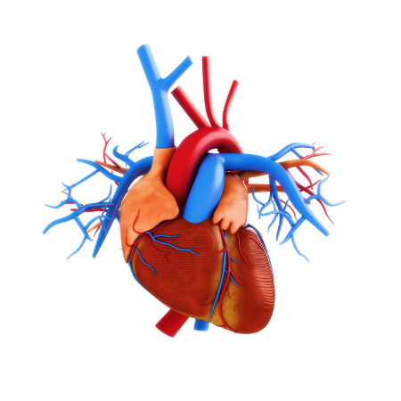
Mitral valve prolapse (aka primary form of myxomatous degeneration of the mitral valve aka floppy mitral valve syndrome) is a valvular heart disease characterized by the displacement of an abnormally thickened mitral valve leaflet into the left atrium during systole. There are various types of MVP, broadly classified as classic and nonclassic. In its nonclassic form, MVP carries a low risk of complications. In severe cases of classic MVP, complications include mitral regurgitation,infective endocarditis, congestive heart failure, and, in rare circumstances, cardiac arrest, usually resulting in sudden death.
The diagnosis of MVP depends upon echocardiography, which uses ultrasound to visualize the mitral valve. The prevalence of MVP is estimated at 2-3% of the population.
The condition was first described by John Brereton Barlow in 1966. In consequence, it may also be referred to as Barlow's Syndrome, and was subsequently termed mitral valve prolapse by J. Michael Criley.
Diagnosis
Echocardiography is the most useful method of diagnosing a prolapsed mitral valve. Two- and three-dimensional echocardiography are particularly valuable as they allow visualization of the mitral leaflets relative to the mitral annulus. This allows measurement of the leaflet thickness and their displacement relative to the annulus. Thickening of the mitral leaflets >5 mm and leaflet displacement >2 mm indicates classic mitral valve prolapse.
Signs and symptoms
Murmur
Upon auscultation of an individual with mitral valve prolapse, a mid-systolic click, followed by a late systolic murmur heard best at the apex is common.
In contrast to most other heart murmurs, the murmur of mitral valve prolapse is accentuated by standing and valsalva maneuver (earlier systolic click and longer murmur) and diminished with squatting (later systolic click and shorter murmur). The only other heart murmur that follows this pattern is the murmur of hypertrophic cardiomyopathy. A MVP murmur can be distinguished from a hypertrophic cardiomyopathy murmur by 1) the presence of a mid-systolic click which is virtually diagnostic of MVP, and 2) the fact that hand grip maneuver intensifies the murmur of MVP and diminishes the murmur of hypertrophic cardiomyopathy. The hand grip maneuver also diminishes the duration of the murmur and delays the timing of the mid-systolic click.[ Hand grip maneuver increases total peripheral resistance (afterload) and therefore increases back pressure on the mitral valve resulting in a more intense murmur.
Both valsalva maneuver and standing decrease venous return to the heart thereby decreasing left ventricular diastolic filling (preload) and causing more laxity on the chordae tendineae. This allows the mitral valve to prolapse earlier in systole, leading to an earlier systolic click (i.e. closer toS1), and a longer murmur.
Treatment
Individuals with mitral valve prolapse, particularly those without symptoms, often require no treatment. Those with mitral valve prolapse and symptoms of dysautonomia (palpitations, chest pain) may benefit from beta-blockers (e.g., propranolol). Patients with prior stroke and/or atrial fibrillation may require blood thinners, such as aspirin or warfarin. In rare instances when mitral valve prolapse is associated with severe mitral regurgitation, mitral valve repair or surgical replacement may be necessary. Mitral valve repair is generally considered preferable to replacement. Current ACC/AHA guidelines promote repair of mitral valve in patients before symptoms of heart failure develop. Symptomatic patients, those with evidence of diminished left ventricular function, or those with left ventricular dilatation need urgent attention.
Prevention of infective endocarditis
Individuals with MVP are at higher risk of bacterial infection of the heart, called infective endocarditis. This risk is approximately three- to eightfold the risk of infective endocarditis in the general population. Until 2007, the American Heart Associationrecommended prescribing antibiotics before invasive procedures, including those in dental surgery. Thereafter, they concluded that "prophylaxis for dental procedures should be recommended only for patients with underlying cardiac conditions associated with the highest risk of adverse outcome from infective endocarditis."
Many organisms responsible for endocarditis are slow-growing and may not be easily identified on routine blood cultures (these fastidious organisms require special culture media to grow). These include the HACEK organisms, which are part of the normal oropharyngeal flora and are responsible for perhaps 5 to 10% of infective endocarditis affecting native valves. It is important when considering endocarditis to keep these organisms in mind.

