-
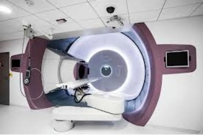
Proton therapy is a type of particle therapy which uses a beam of protons to irradiate diseased tissue, most often in the treatment of cancer. The chief advantage of proton therapy is the ability to more precisely localize the radiation dosage when compared with other types of external beam radiotherapy.
Application
The types of treatments for which protons are used can be separated into two broad categories. The first are those for disease sites that favor the delivery of higher doses of radiation, i.e. dose escalation. In some instances dose escalation has been shown to achieve a higher probability of "cure" (i.e. local control) than conventional radiotherapy.These include (but are not limited to) uveal melanoma (ocular tumors), skull base and paraspinal tumors (chondrosarcoma and chordoma), and unresectable sarcomas. In all these cases proton therapy achieves significant improvements in the probability of local control over conventional radiotherapy.
The second broad class are those treatments where the increased precision of proton therapy is used to reduce unwanted side effects, by limiting the dose to normal tissue. In these cases the tumor dose is the same as that used in conventional therapy, and thus there is no expectation of an increased probability of curing the disease. Instead, the emphasis is on the reduction of the integral dose to normal tissue, and thus a reduction of unwanted effects. prominent examples are pediatric neoplasms (such as medulloblastoma) and prostate cancer. In the case of pediatric treatments there is convincing clinical data showing the advantage of sparing developing organs by using protons, and the resulting reduction of long term damage to the surviving child.
In the case of prostate cancer the issue is not so clear. Some published studies found a reduction in long term rectal and genitio-urinary damage when treating with protons rather than photons (also known as X-ray or gamma ray therapy). Others showed the difference is small, and limited to cases where the prostate is particularly close to certain anatomical structures. The relatively small improvement found may be the result of inconsistent patient set-up and internal organ movement during treatment, which offsets most of the advantage due to increased precision.One source suggests that dose errors around 20% can result from motion errors of just 2.5 mm and another that prostate motion is between 5–10 mm.
Proton, or more generally, hadron therapy of tissue close to the eye affords sophisticated methods to assess the alignment of the eye that can vary significantly from other patient position verification approaches in image guided particle therapy. Position verification and correction have to ensure that sensitive tissue like the optic nerve is spared from the radiation in order to preserve the patient’s vision.
Comparison with other treatment options
The issue of when, whether, and how best to apply this technology is controversial. As of 2012 there have been no controlled trials to demonstrate that proton therapy yields improved survival, or other clinical outcomes (including impotence in prostate cancer) compared to other types of radiation therapy, although a 5-year study of prostate cancer is underway at Massachusetts General Hospital. Proton therapy is far more expensive than conventional therapy.X-ray radiotherapy
Irradiation of nasopharyngeal carcinoma by photon (X-ray) therapy (left) and proton therapy (right)
The figure at the right of the page shows how beams of x-rays (IMRT) left frame and beams of protons right frame, of different energies, penetrate human tissue. A tumor with a sizable thickness is covered by the IMPT spread out Bragg peak (SOBP) shown as the red lined distribution in the figure. The SOBP is an overlap of several pristine Bragg peaks (blue lines) at staggered depths.
Megavoltage X-ray therapy may be described as having more "skin sparing potential" than proton therapy: x-ray radiation at the skin and at very small depths is lower than for proton therapy. One study estimates that passively scattered proton fields have a slightly higher entrance dose at the skin (~75%) compared to therapeutic megavoltage (MeV) photon beams (~60%).X-ray radiation dose falls off gradually, causing unnecessary damage to tissue deeper in the body and damaging the skin and surface tissue opposite the beam entrance. The X-ray advantage of reduced damage to skin at the entrance is partially counteracted by damage to skin at the exit point. Since X-ray treatments are usually done with multiple exposures from opposite sides, each section of skin will be exposed to both entering and exiting X-rays. In proton therapy, skin exposure at the entrance point is higher, but tissues on the opposite side of the body than the tumor receive no radiation. Thus, x-ray therapy causes slightly less damage to the skin and surface tissues, and proton therapy causes less damage to deeper tissues in front of and beyond the target.Surgery
The decision to use surgery or proton therapy (or in fact any radiation therapy) is based on the tumor type, stage, and location. In some instances surgery is superior (e.g. cutaneous melanoma), in some instances radiation is superior (e.g. skull base chondrosarcoma), and in some instances they are comparable (e.g. prostate cancer). In some instances, they are used together (e.g. rectal cancer or early stage breast cancer). The benefit of external beam proton radiation lies in the dosimetric difference from external beam x-ray radiation and brachytherapy in cases, where the use of radiation therapy is already indicated, rather than as a direct competition with surgery.In point of fact, however, in the case of prostate cancer, the most common indication for proton beam therapy, no clinical study directly comparing proton therapy to surgery, brachytherapy, or other treatments has even shown any clinical benefit for proton beam therapy. Indeed, the largest study to date showed greater rectal complications and more erectile dysfunction following proton beam treatment compared to conventional radiation therapy.

