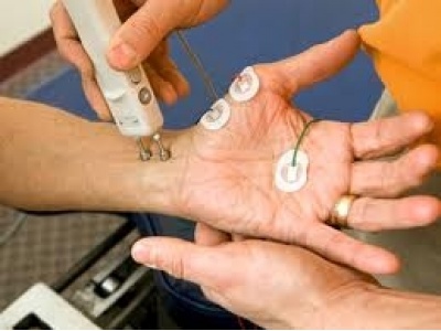
Electromyography (EMG) - a method ofstudy of the neuromuscular system by detecting electrical potentials of muscles.
Elektroneyrography (ENG) - a method ofassessing how quickly the electrical signalcarried by nerves.
Determination of the electrical activity ofmuscles and nerves helps identify diseases in which muscle tissue pathology observed(eg, muscular dystrophy) or neural tissue(amyotrophic lateral sclerosis, or peripheral neuropathy). For completeness of the survey, both these methods of research -and EMG and ENG - held together.
Indications for EMG
pathology from the muscle and nervous tissue, as well as the junction of nerve and muscle (neuromuscularsynapse) herniated disc, amyotrophic lateral sclerosis, myasthenia gravis.
Determine the cause of Violations of the muscles, nerves, spinal cord or of the brain, which can cause such changes as weakness, paralysis or muscle twitching.
EMG does not reveal pathology of the spinal cord or brain.
Indications for EWS:
pathology of the peripheral nervous system, which includes all the nerves that leave the spinal cord and brain.The nerve conduction studies of the electrical signal is often used to diagnose carpal tunnel syndrome andGuillain-Barre syndrome.
Possible complications
EMN and ENG - safe methods.
There are several types of electromyography:
Interference EMG assigned cutaneous electrodes at arbitrary reductions in passive muscle or bend thelimbs.
Local EMG. Draining potentials produced by the concentric electrodes immersed in the muscle.
Stimulus EMG (electro-neuromyography). Diversion biopotentials performed as cutaneous and needle electrodes during stimulation of peripheral nerve.
Furthermore, there is the so-called external sphincter electromyography to determine the electrical activity of the external sphincter of the bladder. Moreover, its activity may be defined as using needle electrodes, and throughthe anal and cutaneous.

