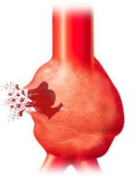
Aortic dissection occurs when a tear in the inner wall of the aorta causes blood to flow between the layers of the wall of the aorta, forcing the layers apart. In most cases this is associated with severe characteristic chest or abdominal paindescribed as "tearing" in character, and often with other symptoms that result from decreased blood supply to other organs. Aortic dissection is a medical emergency and can quickly lead to death, even with optimal treatment, as a result of decreased blood supply to other organs, cardiac failure, and sometimes rupture of the aorta. Aortic dissection is more common in those with a history of high blood pressure, a known thoracic aortic aneurysm, and in a number of conditions that affect blood vessel wall integrity such as Marfan syndrome and the vascular subtype of Ehlers–Danlos syndrome. The diagnosis is made with medical imaging (computed tomography, magnetic resonance imaging or echocardiography).
The treatment of aortic dissection depends on the part of the aorta involved. Surgery is usually required for dissections that involve the aortic arch, while dissections of the part further away from the heart may be treated with blood pressure lowering only. Since the 1990s endovascular aneurysm repair (carried out from inside the blood vessels) has been used in specific cases.
Aortic dissection is relatively rare, occurring at an estimated rate of 2–3.5 per 100,000 people every year. It is more common in males. Mean age at diagnosis is 63, although all age groups may be affected. Many cases of aortic dissection (40%) lead to death so rapidly that the person doesn't make it to a hospital in time. The first case of aortic dissection described was in the post-mortem examination of King George II of Great Britain in 1760. Surgery for aortic dissection was introduced in the 1950s.
Signs and symptoms
About 96% of individuals with aortic dissection present with severe pain that had a sudden onset. It may be described as tearing, stabbing, or sharp in character. 17% of individuals will feel the pain migrate as the dissection extends down the aorta. The location of pain is associated with the location of the dissection. Anterior chest pain is associated with dissections involving the ascending aorta, while interscapular (back) pain is associated with descending aortic dissections. If the pain is pleuritic in nature, it may suggest acute pericarditis caused by hemorrhage into the pericardial sac. This is a particularly dangerous eventuality, suggesting that acute pericardial tamponade may be imminent. Pericardial tamponade is the most common cause of death from aortic dissection.
While the pain may be confused with the pain of a myocardial infarction (heart attack), aortic dissection is usually not associated with the other signs that suggest myocardial infarction, including heart failure and ECG changes.
Individuals with aortic dissection who do not present with pain have chronic dissection.
Less common symptoms that may be seen in the setting of aortic dissection include congestive heart failure (7%), syncope (9%), cerebrovascular accident (3-6%), ischemic peripheral neuropathy, paraplegia, cardiac arrest, and sudden death. If the individual had a syncopal episode, about half the time it is due to hemorrhage into the pericardium leading to pericardial tamponade.
Neurologic complications of aortic dissection (i.e., cerebrovascular accident (CVA) and paralysis) are due to involvement of one or more arteries supplying portions of the central nervous system.
If the aortic dissection involves the abdominal aorta, compromise of the branches of the abdominal aorta is possible. In abdominal aortic dissections, compromise of one or both renal arteries occurs in 5–8% of cases, while mesenteric ischemia (ischemia of the large intestines) occurs 3–5% of the time.
Blood pressure
While some patients with an aortic dissection have a history of hypertension, the blood pressure is quite variable at presentation with acute aortic dissection, and tends to be higher in individuals with a distal dissection. In individuals with a proximal aortic dissection, 36% present withhypertension, while 25% present with hypotension. In those who present with distal aortic dissections, 70% present with hypertension while 4% present with hypotension.
Severe hypotension at presentation is a grave prognostic indicator. It is usually associated with pericardial tamponade, severe aortic insufficiency, or rupture of the aorta. Accurate measurement of the blood pressure is important. Pseudohypotension (falsely low blood pressure measurement) may occur due to involvement of the brachiocephalic artery (supplying the right arm) or the left subclavian artery (supplying the left arm).
Aortic insufficiency
Aortic insufficiency (AI) occurs in half to two-thirds of ascending aortic dissections, and the murmur of aortic insufficiency is audible in about 32% of proximal dissections. The intensity (loudness) of the murmur is dependent on the blood pressure and may be inaudible in the event of hypotension.
There are multiple etiologies for AI in the setting of ascending aortic dissection. The dissection may dilate the annulus of the aortic valve, so that the leaflets of the valve cannot coapt. Another mechanism is that the dissection may extend into the aortic root and detach the aortic valve leaflets. The third mechanism is that if there was an extensive intimal tear, the intimal flap may prolapse into the LV outflow tract, causing intimal intussusception into the aortic valve preventing proper valve closure.
Myocardial infarction
Myocardial infarction (heart attack) occurs in 1–2% of aortic dissections. The etiology of the infarction is involvement of the coronary arteries (the arteries that supply the heart) in the dissection. The right coronary artery is involved more commonly than the left coronary artery. If the myocardial infarction is treated with thrombolytic therapy, the mortality increases to over 70%, mostly due to hemorrhage into the pericardial sac causing pericardial tamponade.
Because aortic dissection may present to the emergency room physician similar to a myocardial infarction, the physician must be careful to make the proper diagnosis prior to initiating treatment for myocardial infarction, since the treatment regimen for myocardial infarction can be lethal to an individual presenting with aortic dissection.
Pleural effusion
A pleural effusion (fluid collection in the space between the lungs and the chest wall or diaphragm) can be due to either blood from a transient rupture of the aorta or fluid due to an inflammatory reaction around the aorta. If a pleural effusion were to develop due to an aortic dissection, it is more commonly in the left hemithorax rather than the right hemithorax.
Diagnosis
Because of the varying symptoms and signs of aortic dissection depending on the initial intimal tear and the extent of the dissection, the proper diagnosis is sometimes difficult to make.
While taking a good history from the individual may be strongly suggestive of an aortic dissection, the diagnosis cannot always be made by history and physical signs alone. Often the diagnosis is made by visualization of the intimal flap on a diagnostic imaging test. Common tests used to diagnose an aortic dissection include a CT scan of the chest with iodinated contrast material and a trans-esophageal echocardiogram. The proximity of the aorta to the esophagus allows the use ofhigher-frequency ultrasound for better anatomic images. Other tests that may be used include an aortogram or magnetic resonance angiogram (MRA) of the aorta. Each of these tests have varying pros and cons and they do not have equal sensitivities and specificities in the diagnosis of aortic dissection.
In general, the imaging technique chosen is based on the pre-test likelihood of the diagnosis, availability of the testing modality, patient stability, and the sensitivity and specificity of the test.
D-dimer
A blood D-dimer level less than 500 ng/ml may be able to rule out the diagnosis of aortic dissection alleviating the need for further imaging.
Chest X-ray
.
Widening of the mediastinum on an x-ray of the chest has moderate sensitivity in the setting of an ascending aortic dissection. However, it has low specificity, as many other conditions can cause a widening of the mediastinum on chest x-ray.
The calcium sign is a finding on chest x-ray that suggests aortic dissection. It is the separation of the intimal calcification from the outer aortic soft tissue border by 10 mm.
Pleural effusions may be seen on chest x-ray. They are more commonly seen in descending aortic dissections. If seen, they are typically in the left hemithorax.
Other findings include obliteration of the aortic knob, depression of the left mainstem bronchus, loss of the paratracheal stripe, and tracheal deviation.
About 12 to 20% of individuals presenting with an aortic dissection have a "normal" chest x-ray; therefore, a normal chest radiograph does NOT rule out aortic dissection. If the clinical index of suspicion is high, it is imperative to rule out dissection with another imaging modality (CT angiogram, MRA, aortogram, or transesophageal echo)
Computed tomography
Computed tomography angiography is a fast non-invasive test that will give an accurate three-dimensional view of the aorta. These images are produced by taking rapid thin cut slices of the chest and abdomen, and combining them in the computer to create cross-sectional slices. In order to delineate the aorta to the accuracy necessary to make the proper diagnosis, an iodinated contrast material is injected into a peripheral vein. Contrast is injected and the scan performed using a Bolus Tracking method. This is a type of scan timed to an injection to capture the contrast as it enters the aorta. The scan will then follow the contrast as it flows though the vessel.
It has a sensitivity of 96 to 100% and a specificity of 96 to 100%. Disadvantages include the need for iodinated contrast material and the inability to diagnose the site of the intimal tear.
MRI
Magnetic resonance imaging (MRI) is currently the gold standard test for the detection and assessment of aortic dissection, with a sensitivity of 98% and a specificity of 98%. An MRI examination of the aorta will produce a three-dimensional reconstruction of the aorta, allowing the physician to determine the location of the intimal tear, the involvement of branch vessels, and locate any secondary tears. It is a non-invasive test, does not require the use of iodinated contrast material, and can detect and quantitate the degree of aortic insufficiency.
The disadvantage of the MRI scan in the face of aortic dissection is that it has limited availability and is often located only in the larger hospitals, and the scan is relatively time consuming. Due to the high intensity magnetic fields used during MRI, an MRI scan is contraindicated in individuals with metallic implants. In addition, many individuals experience claustrophobia while in the MRI scanning tube.
Transesophageal echocardiography
The transesophageal echocardiogram (TEE) is a relatively good test in the diagnosis of aortic dissection, with a sensitivity of up to 98% and a specificity of up to 97%. It has become the preferred imaging modality for suspected aortic dissection. It is a relatively non-invasive test, requiring the individual to swallow the echocardiography probe. It is especially good in the evaluation of AI in the setting of ascending aortic dissection, and to determine whether the ostia (origins) of the coronary arteries are involved. While many institutions give sedation during transesophageal echocardiography for added patient comfort, it can be performed in cooperative individuals without the use of sedation. Disadvantages of the TEE include the inability to visualize the distal ascending aorta (the beginning of the aortic arch), and the descending abdominal aorta that lies below the stomach. A TEE may be technically difficult to perform in individuals with esophageal strictures or varices.